Main Features Of Multiple Pilomatricoma
Multiple pilomatricoma is usually associated with Steirnet’s disease or myionotic dystrophy or a family history of lesions of this type. It can also be related to other diseases.

Multiple pilomatricoma is a fairly rare lesion. In general, pilomatricoma is a benign tumor that arises in the cells of the hair matrix and that usually occurs alone. The multiple type is much more rare.
There are no conclusive data on the incidence of multiple pilomatricoma. There are already large gaps in the data about pilomatricoma in its typical form, because it is frequently confused with other pathologies.
Despite the above, what is known is that multiple pilomatricoma represents between 2% and 3.5% of all pilomatricoma cases. They take place mainly when there is a family history of this type of injury.
What is multiple pilomatricoma

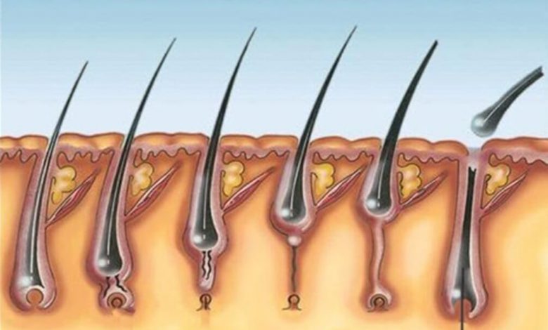
Let’s first define pilomatricoma as such. This is a benign tumor that arises in the matrix of the hair follicle and whose cause is unknown. It occurs mainly during childhood. It is estimated that it is equivalent to 10% of skin tumors that appear in children.
These types of tumors are located in the deep dermis and frequently extend to fat. When there are several tumors of this type, it is called multiple pilomatricoma. These cases are usually associated with heredity or concomitant diseases.
So far only 40 cases of multiple pilomatricoma related to inheritance and 30 cases associated with Steirnet’s disease or myionotic dystrophy have been described. In fact, for some authors, multiple pilomatricoma is only one symptom of such myionotic dystrophy.
Cases of other associated diseases have also been found, such as Gardner syndrome, Rubinstein-Taybi syndrome, medullary thyroid carcinoma, trisomy, mild coagulation disorders, and sternal closure defects. Likewise, there are patients without associated anomalies.
Characteristics

Pilomatricoma presents as a hard nodule, usually the same color as the skin. It can appear at any age, but 60% of cases appear in the first decade of life. They are also slightly more common in the sixth and seventh decades of life. It slightly predominates in the female gender.
In more than half of the cases these nodules appear on the head and neck, but they can appear anywhere on the body that has hair follicles. It has a bluish-reddish color. When you feel it, you can feel the stony interior and the embossed surface.
Multiple pilomatricoma can appear before or after myotonic dystrophy or Steirnet’s disease. This is a genetically transmitted muscular dystrophy characterized by the progressive deterioration of voluntary muscles. It manifests itself more severely and quickly in each next generation.
In multiple pilomatricoma there are several lesions that sometimes converge together and form a kind of plaque. The growth of the nodules is slow and asymptomatic. Generally, it is the same patient who notices its presence when palpating.
Causes and diagnosis
A complex of proteins called catenins is thought to be involved in multiple pilomatricoma . These are responsible for joining the actin of the cytoskeleton with cadherins. Everything indicates that the final cause of this problem is the beta-catenin mutation, which has been verified in 75% of pilomatricomas.
The clinic of this disease is characterized by myotonia, which is the difficulty to relax the muscles. There is also muscle weakness and atrophy, as well as lens opacities, moderate mental retardation, early frontal alopecia, testicular atrophy, heart block, and electromyogram abnormalities.
The diagnostic is fundamentally clinical. Sometimes complementary studies are done to confirm this diagnosis, such as ultrasound. Interrogation, hemogram, and urine and stool tests can confirm or rule out the presence of associated syndromes.
Other data of interest
Sometimes the appearance of the nodules is fine, atrophic and wrinkled or pseudo-bullous. This occurs in only 2% of cases, often in young women with limb injuries.
Only very rarely do tumors have a crusty appearance, similar to that of an ulcer. Only 11 such cases are known to exist. In these cases we speak of perforating tumors.
Simple pilomatricoma and multiple pilomatricoma tumors are identical. However, there are four cases in which multiple tumors present with macular atrophy or anetoderma. Long-term follow-up should be maintained when confirming the presence of multiple pilomatricoma.